Neurotechniques, also known as neuroscience techniques, are various methods and tools used in the field of neuroscience to study the structure, function, and mechanisms of the nervous system. These techniques enable researchers to investigate the brain and its activities at various levels, ranging from molecular and cellular to systems and behavioral levels. Neurotechniques play a crucial role in advancing our understanding of the brain and its role in various physiological and psychological processes. Here are some common neurotechniques used in neuroscience research:
⦁ Electroencephalography (EEG): EEG is a non-invasive technique that records the electrical activity of the brain using electrodes placed on the scalp. It is often used to study brain waves and patterns associated with different states of consciousness, such as sleep stages, cognitive processes, and neurological disorders.
⦁ Functional Magnetic Resonance Imaging (fMRI): fMRI is a non-invasive imaging technique that measures changes in blood flow and oxygenation to infer neural activity in different brain regions. It allows researchers to study brain function and connectivity during various cognitive tasks and emotional experiences.
⦁ Positron Emission Tomography (PET): PET is a nuclear medicine imaging technique that uses radioactive tracers to monitor metabolic and biochemical processes in the brain. It is commonly used to study brain activity, neurotransmitter systems, and neuroreceptor binding.
⦁ Transcranial Magnetic Stimulation (TMS): TMS is a non-invasive neurostimulation technique that uses magnetic fields to stimulate specific brain areas. It is used to investigate brain function, map cortical regions, and study the effects of brain stimulation on cognition and behavior.
⦁ Patch-Clamp Electrophysiology: This technique involves placing a microelectrode on the cell membrane of neurons to record and manipulate their electrical activity. It allows researchers to study individual neurons and investigate synaptic transmission and ion channel function.
⦁ Optogenetics: Optogenetics is a method that uses light to control the activity of specific neurons genetically modified to express light-sensitive proteins. It allows researchers to selectively activate or inhibit neural circuits, providing insights into brain function and behavior.
⦁ Calcium Imaging: This technique involves using fluorescent indicators to monitor changes in intracellular calcium levels, which are associated with neuronal activity. It helps researchers study neural network activity in real-time.
⦁ Neuropsychological Testing: Neuropsychological tests are behavioral assessments used to evaluate cognitive functions in individuals with brain injuries or neurological disorders. These tests provide valuable information about brain-behavior relationships.
⦁ Single-Unit Recording: Invasive single-unit recording involves placing an electrode directly into the brain to record the electrical activity of individual neurons. It is used in animal research to study neuronal firing patterns and coding of sensory information.
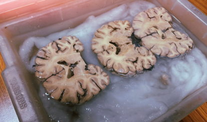
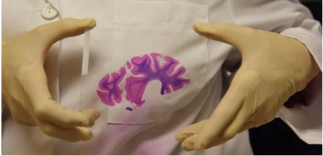
⦁ Histology: Histology involves the study of tissues at a microscopic level. In neuroscience, researchers use histological techniques to examine the cellular composition and organization of brain tissue. Common histological staining methods include Hematoxylin and Eosin (H&E) staining, Nissl staining, and Golgi staining, which allow visualization of different cell types and neural structures.
⦁ Immunohistochemistry (IHC): IHC is a technique used to visualize specific proteins or antigens within brain tissue. Antibodies labeled with fluorescent or chromogenic markers are used to bind to target proteins, enabling their detection and localization in neurons or other brain cells.
⦁ In Situ Hybridization (ISH): ISH is a molecular biology technique used to detect the presence and distribution of specific RNA molecules within brain tissue. It allows researchers to study the expression patterns of genes in different brain regions.
⦁ Tracing Techniques: Tracing techniques involve the injection of tracers, such as fluorescent dyes or viral vectors, into specific brain regions. These tracers label and trace neuronal pathways, providing information about neural connectivity and circuitry.
⦁ Electron Microscopy (EM): EM is a high-resolution imaging technique that allows researchers to study ultrastructural details of neurons and synapses. It provides information about the cellular organelles and synaptic morphology in the brain.
⦁ Neuroanatomical Imaging: Advanced imaging techniques, such as diffusion tensor imaging (DTI) and structural magnetic resonance imaging (MRI), are used to study the macroscopic structure and connectivity of the brain. These techniques provide detailed maps of white matter tracts and brain regions.
⦁ Molecular Biology Techniques: Molecular biology techniques are used to study the genetic and molecular processes in the brain. Polymerase chain reaction (PCR), Western blotting, and gene expression analysis are common techniques used to study gene expression and protein levels in the brain.
⦁ Next-Generation Sequencing (NGS): NGS is a powerful genomic technique used to analyze large-scale DNA and RNA sequencing data. It allows researchers to study the transcriptome and epigenome of the brain, providing insights into gene regulation and expression.
⦁ Patch-Clamp Electrophysiology: While mentioned before, patch-clamp electrophysiology is also a valuable molecular technique used to study the electrical properties of individual neurons and their ion channel function.
In the fascinating field of neuroscience, researchers have access to a diverse array of neurotechniques, each offering unique advantages and limitations. From histological and anatomical methods to molecular biology techniques, these tools provide valuable insights into the intricate workings of the brain and its profound impact on behavior and cognition. By combining and harnessing the power of these neurotechniques, scientists can gain a comprehensive understanding of the brain’s structure, function, and molecular processes. This integrated approach plays a pivotal role in unraveling the underlying mechanisms of various neurological disorders, understanding brain development, and exploring the intricacies of cognitive processes. The continuous integration of histological, anatomical, and molecular approaches in neuroscience research has resulted in remarkable advancements in our knowledge of the brain’s complexities, shaping the path towards groundbreaking discoveries in this ever-evolving scientific frontier.
Explore Related Articles